
Reviewing a peripheral blood smear requires active searching and documentation of morphological characteristics of red and white blood cells. In addition to quantity, size, and shape, the laboratory scientist must identify unusual cellular inclusions observed on the slide, and note them on the laboratory report.
When a laboratory scientist finds these things, they call over everyone in the lab to see the exciting and unusual find. Then they look for more of them. It’s rewarding to find these things. Like a game of hide and seek.
When they find these exciting and clinically relevant items, they know it will have an effect on the patient’s diagnosis and treatment. They receive an immediate internal reward for finding something cool and for knowing they are performing meaningful work on behalf of the patients. They are satisfying their inner science nerd!
RBC inclusions such as basophilic stippling, Pappenheimer bodies, Heinz Bodies (indicated by bite cells on the Wright-Giemsa stain), Howell-Jolly bodies, Cabot rings, hemoglobin C crystals, and hemoglobin SC crystals may be seen.
WBC inclusions commonly seen are Dohle bodies, Auer rods, and toxic granulation.
Students new to Hematology will appreciate this summary of the inclusions, their composition, disease correlation, and stains used to visualize them. Scientists who have been performing differentials much longer than I have, I appreciate any feedback you might have!
Red Blood Cell Inclusions
Basophilic Stippling
Students confuse basophilic stippling with toxic granulation when first learning blood cell morphology. Basophilic stippling occurs in red blood cells while toxic granulation occurs in white blood cells.
Basophilic means it appears blue/purple on Wright-Giemsa stain. Stippling means there are dots spread across the entire red blood cell. The stippling can be fine and barely visible, or it can be coarse and large.
- Stain: Wright-Giemsa
- Composition: mitochondria and RNA remnants caused by toxic conditions affecting hemoglobin synthesis
- Disease correlation: Lead poisoning, malignant conditions, alcoholism, or any condition affecting heme synthesis, heme being the iron-containing building block for hemoglobin, and hemoglobin being the substance inside red blood cells that binds oxygen and carries it throughout the body
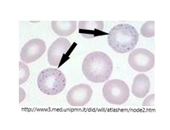
Pappenheimer Bodies
Pappenheimer bodies can be confused for basophilic stippling if a large number are present within a red cell, such as the cell indicated by the left arrow. This image is a Wright-stained blood smear.
Differentiation between the two inclusions is done using Prussian Blue stain, which will stain the iron in Pappenheimer bodies, and will not stain basophilic stippling because they lack iron. When using Prussian Blue stain, the inclusions are called siderotic granules.
- Stain: Wright-Giemsa (Pappenheimer bodies), Prussian Blue (siderotic granules)
- Composition: Iron
- Disease correlation: sideroblastic anemia, thalassemia, alcoholism; ineffective red cell development (erythropoiesis)
Howell-Jolly Bodies
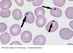
Usually, Howell-Jolly or HJ Bodies occur as singlets within red cells, but can have multiples in some instances. They can be confused with Pappenheimer bodies when multiples are present. They may be distinguished from Pappenheimer bodies by using Prussian Blue stain. They are more prevalent after a patient has their spleen removed.
- Stain: Wright-Giemsa
- Composition: DNA remnants from nuclear expulsion. Normally removed by the spleen.
- Disease correlation: hyposplenism or post-splenectomy; abnormal hemoglobin diseases; pernicious anemia (Vitamin B12 deficiency).
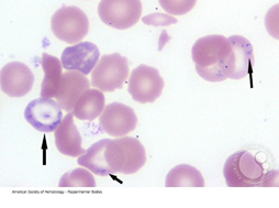
Heinz Bodies (associated with bite cells)
Students, make sure you take note of what stain is used for the images being shown. The image on top is peripheral blood stained with a supravital stain such as New Methylene Blue. Heinz bodies will not stain on Wright-Giemsa, and will stain with a supravital stain.
Make sure you don’t mistake Howell-Jolly bodies for Heinz bodies when performing your peripheral smear evaluation. Routine peripheral smears are stained with Wright-Giemsa, and Heinz bodies will not show.
Heinz bodies tend to bulge from the edge of the red cell membrane. I like to think of Heinz ketchup to help me differentiate them from HJ bodies. The Heinz bodies are on the edge waiting to be dipped into the Heinz ketchup, but the HJ bodies are not.
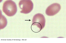
The image on the bottom is Wright-Giemsa stain, showing a bite cell. It looks like a bite was taken out of it. This occurs when the spleen removes the Heinz body as the red cell passes through it. The membrane is damaged by the removal, but it does not destroy the red cell. The membrane is very flexible and resilient. Taking only part of it allows the red cell to survive and continue transporting oxygen through the body.
- Stain: Supravital, such as New Methylene Blue
- Composition: denatured hemoglobin precipitate due to defective red cell metabolism in the hexose monophosphate shunt
- Disease correlation: G6PD deficiency, oxidant drugs, infections
Cabot Rings
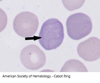
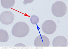
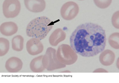
Cabot rings are rarely seen during peripheral smear evaluation. They are easy to miss, being thin rings, figure 8’s, or letter B’s inside the red cell. They may be dismissed as artifact by the novice laboratory scientists.
They are thought to be remnants of mitotic spindle tubules after the red cell nucleus as been expelled.
They can be present along with other things such as basophilic stippling or polychromasia (when the red cell has a bluer appearance because it is still young).
- Stain: Wright-Giemsa
- Composition: Mitotic spindle microtubules
- Disease correlation: conditions of dyserythropoiesis (abnormal red cell production) such as lead poisoning, megaloblastic anemia, leukemia, and myelodysplastic syndrome
White Blood Cell Inclusions
Toxic Granulation
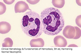
Toxic granulation is seen in neutrophil leukocytes.
I’ve included an image or 2 normal neutrophils first for comparison. Notice that although they have granules in the cytoplasm, the granules are more pink in color.
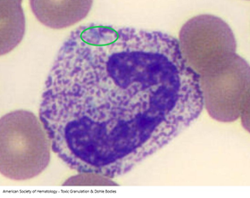
The image on the bottom is a neutrophil, possibly a band neutrophil, with uniform toxic granulation throughout the cytoplasm. Notice that the granules are more purple in color. The granules can be very fine, or very course. These granules are somewhere in the middle.
Also notice the green circle. Under all that toxic granulation are several Dohle bodies, which are pale blue.
Generally, toxic granulation and Dohle bodies are not disease specific. They are reactive changes to a number of conditions that stress the body.
- Stain: Wright-Giemsa
- Composition: enzymes – peroxidases and acid hydrolases
- Disease correlation: nonspecific; may be seen in severe bacterial infections, vasculitis, chemotherapy, or toxemia of pregnancy
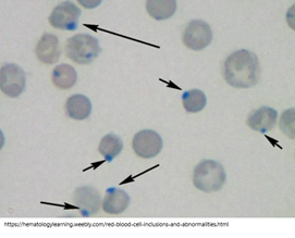
Dohle Bodies
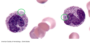
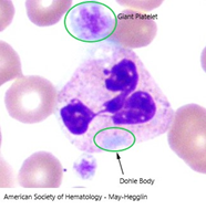
Dohle bodies are pale blue oval inclusions in neutrophil leukocytes measuring 1-3 microns in diameter. They may appear as singlets or multiples, and may be hard to see through toxic granulation. Most circles on these images indicate the Dohle body location.
Dohle bodies come from RNA and are generally not disease specific, as mentioned above.
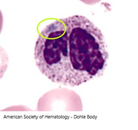
The image of May-Hegglin anomaly shows a Dohle body not obscured by toxic granulation. Note with this anomaly, you will also see giant platelets along with the Dohle bodies. May-Hegglin is a genetic disorder, which is why toxic granulation is typically absent.
- Stain: Wright-Giemsa
- Composition: RNA
- Disease correlation: nonspecific; may be seen in severe infections, pregnancy, severe burns, chemotherapy, May-Hegglin anomaly, Chediak-Higashi disorder
Auer Rods
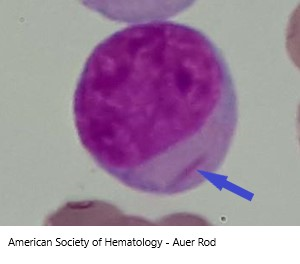
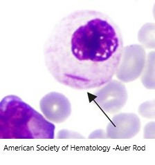
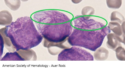
Auer rods are azurophilic, meaning they are reddish in color on Wright-Giemsa stain. They are shaped like needles and may be singlets or multiples.
When present in Acute Myeloid Leukemia or Myelodysplastic Syndromes, they indicate a worse prognosis for the patient.
They result from fusion of primary granules, leading to a lack of or fewer secondary granules being formed.
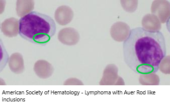
Cases are being reported of Auer rod-like inclusions in lymphoma cells, as shown in the bottom image. This lymphoma was confirmed by flow cytometry as a low-grade B-cell lymphoma.
When these rods are seen in lymphoma cases, it is believed that they are immunoglobulin deposits, which makes sense when you consider that B-cells produce antibodies which are immunoglobulins.
- Stain: Wright-Giemsa
- Composition: fused primary granules
- Disease correlation: Myelodysplastic Syndromes (MDS), Acute Myeloid Leukemia (AML)
In Summary
Red and white cell inclusions are important to report as part of the patient’s peripheral smear evaluation. You may need to submit the slides for review by the Pathologist to confirm if you are unsure of what you are seeing.
Knowing how important it is to identify these rare inclusions drives laboratory scientists to complete thorough slide reviews, and include other scientists in the excitement when one is found.
I encourage you to channel your inner nerd and post images of your exciting finds in the comments of this blog. We’d love to see them! As always, please make sure they do not violate any privacy rules.
Happy hunting!