
What are they?
As laboratory professionals, one laboratory test we perform is a peripheral blood smear evaluation. We place a drop of patient blood on a glass slide, stain it with Wright-Giemsa stain, and look at it under the microscope. We have criteria to determine if the red and white blood cells look normal or abnormal. If abnormal, we can often tell what the disease is based on the shape, size, and color of the cells on the slide.
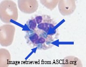
“Inclusions” are possible abnormalities seen inside red and white blood cells. These are things other than the nucleus and cytoplasm that are observed inside the cells.
Blue-green crystals are white blood cell inclusions that have garnered more interest in recent years.
What are they made of?
Blue-green crystals are thought to be made of lipopigments composed of protein, lipid (lipofuscin and ceroid), and a metal such as iron. The crystals will not degrade because peptide crosslinking occurs, forming something similar to plastic.
It is possible the crystals form as a result of lysosomal functions in the cells. Further investigation must occur to confirm this theory.
Death crystals? Really?
Lab scientists have called them “crystals of death”. They are often associated with critically ill patients experiencing severe tissue damage and organ failure, leading to patient death within 2-3 days of the crystals being discovered on the peripheral blood smear.
This may be a misnomer as patients with these white cell inclusions have recovered from their conditions. As laboratory professionals, we should discourage the use of this phrase to avoid negatively influencing the interpretation of the patient’s results.
The misnomer could be caused by bias associated with:
- underreporting these crystals when they are present on the peripheral blood smear, and/or
- increased use of automated instrument differential reports with no slide review.
Not reporting them when they are present skews data obtained from patient records when determining correlation of crystal presence on the smear with specific patient conditions.
Automated instruments may not detect the crystals. If the size and number of the cells is normal, the instruments will call the specimen normal.
Who can get them?
Cases reporting blue-green inclusions have included patients with conditions such as:
- Liver failure
- Kidney disease/failure
- Renal carcinoma
- Heart failure
- COVID-19
- Sepsis
Common links between these conditions include:
- Tissue damage
- Elevated liver enzymes such as aspartate aminotransferase (AST) and alanine aminotransferase (ALT)
- Elevated lactic acid
- Elevated lactate dehydrogenase (LD)
More analysis correlating the presence of blue-green inclusions with other lab tests and associated diseases/conditions is needed to determine the true relationships between them.
Are they important to patient outcome?
As mentioned earlier, these inclusions have been known as blue-green crystals of death due to an apparent correlation with patient death within days of identifying the crystals in the patient’s specimen.
The crystals alone, if not used in conjunction with other lab findings and the patient’s clinical symptoms, may provide a misleading picture of the patient’s condition. Not all patients die within days of the crystals appearing. Some patients recover and the crystal formation disappears in subsequent patient specimens.
High lactic acid levels may be a good predictor of poor prognosis at the time the crystals are seen on the peripheral smear, based on cases reported in the literature. However, not all patients with these inclusions have lactic acid results to correlate. Not all patients with lactic acid results have peripheral blood smear evaluation performed to look for the inclusions. This might be an opportunity for a student researcher to review results and look at peripheral smears to ascertain if there is a true correlation with patient prognosis.
Consult your Pathologist when these crystals are identified on a peripheral smear. The Pathologist can review the patient’s chart along with the peripheral smear and possibly discuss the patient’s results with the physician to arrive at a decision about the importance of including the crystals in the patient record.
As of this writing, no consensus exists in the literature for reporting these blue-green inclusions in the patient report versus not. More data is needed.
Share your photos with us!
If you come across blue-green white cell inclusions as you are looking at your peripheral smears, take a photo and share with us on social media! We are on Facebook, Instagram, and Twitter. Please remember no HIPAA protected information, please, just the photo.
Future blogs will discuss other blood cell inclusions and their association with diseases/conditions. Hope to see more photos there, too!