In current practice, where a large majority of laboratory testing is automated, it is often a nice change of pace to be able to manually review a patient specimen. One area of laboratory medicine that still requires manual review is microscopic urinalysis.
Going into my MLS education, I had a general idea of the basic components of urine sediment, such as white cells, red cells, epithelial cells, and bacteria. But then I discovered something a little less mundane to look at: urine crystals!
So what are these fascinating little shapes that can be seen in urine? Under the right conditions, some of the various chemicals filtered by the kidneys may precipitate into solids. As you will see shortly, pH is a large driving force of crystal precipitation, and oftentimes is used as the first factor to identify the crystal.
Some crystals are clinically insignificant, while others can be an indicator of a genetic abnormality. Let’s take a look at some of the crystals you may see in your career in the lab.
Uric Acid
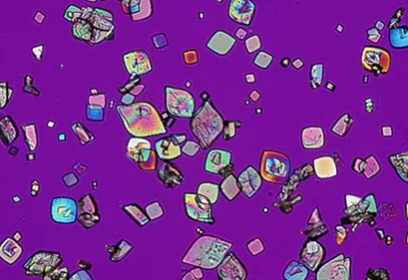
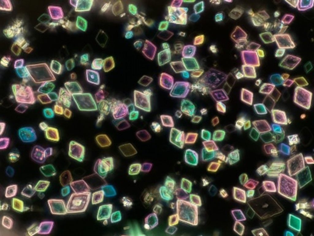
The most common crystal, uric acid, will precipitate into solids in acidic urine (pH ~5.5). Their shape can vary, but most often they will appear as diamonds or rhomboids. In small quantities, uric acid crystals hold little clinical significance other than suggesting acidic urine, a high-protein diet, or slight dehydration. However, large amounts of these crystals in the urine typically point to some underlying cause of hyperuricemia. Some examples are severe dehydration, kidney disease, gout, or they may even be seen after a seizure. An excessive build-up of uric acid in the kidneys may also lead to kidney stone formation.
It should also be noted that just like the uric acid crystals seen in joint fluid from patients with gout, the uric acid crystals in urine will polarize light as well. If you have the opportunity, I definitely recommend examining urine sediment with a polarized microscope!
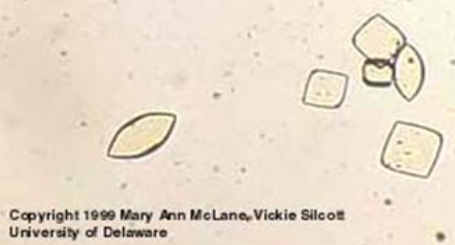
Calcium Oxalate
Like uric acid, calcium oxalate crystals can build up and form kidney stones. These crystals may be seen in urine of various pH levels. They have two forms, but will most often appear in the dihydrate form, with a shape resembling a square envelope. While not typically a clinically significant finding, these crystals can be found in a patient’s urine if they’ve ingested ethylene glycol. Reporting these crystals may signal to the patient’s provider that they are at risk for kidney stones due to insufficient water intake or a diet rich in salt and food containing oxalates.
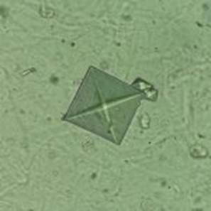
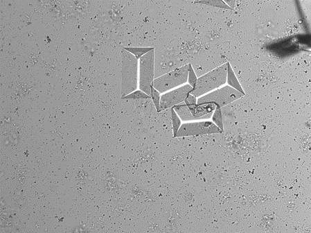
Triple Phosphate
Triple phosphate crystals (also known as magnesium ammonium phosphate or struvite) can be found in alkaline urine. While they may be seen in healthy urine, they usually appear alongside a urinary tract infection (UTI). Bacteria that produce nitrogen-containing compounds will increase the pH of the urine, allowing these crystals to precipitate. Their appearance is typically described as a “coffin lid.”
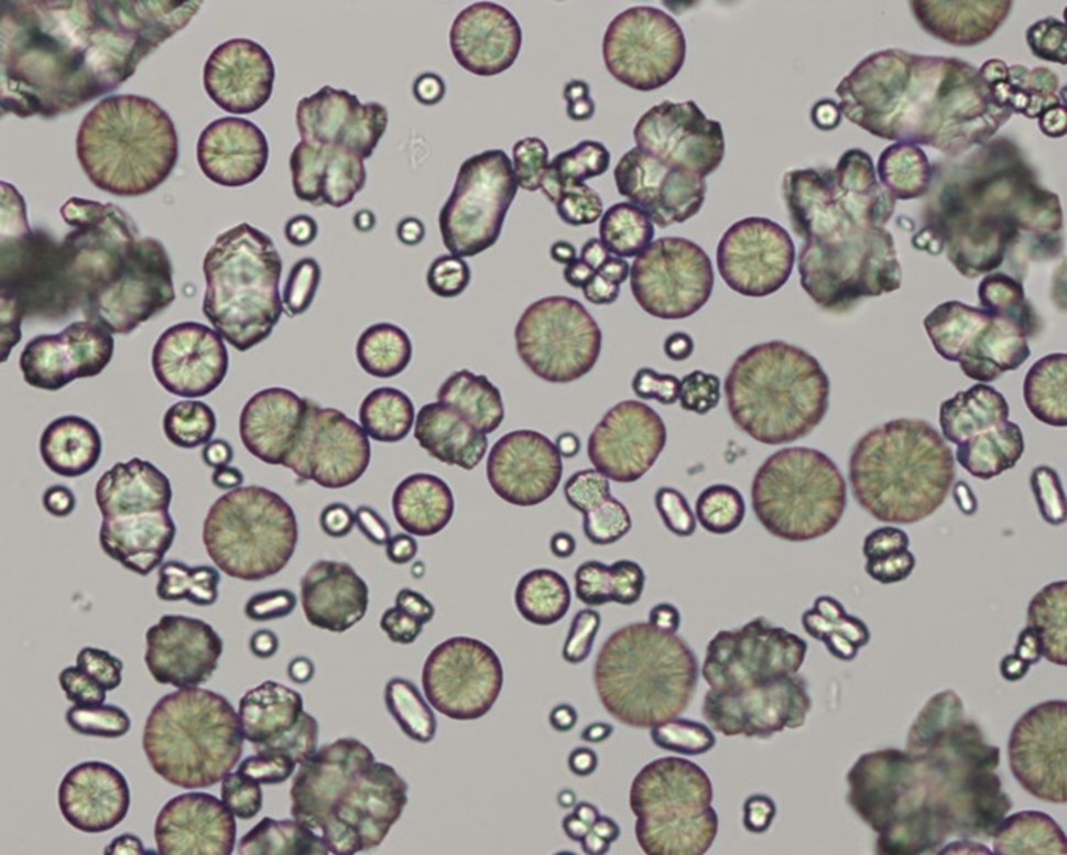
Calcium Carbonate
Calcium carbonate crystals can be found in alkaline urine. Their appearance is not very clinically significant. They may be seen in patients with decreased urine volume, increased calcium levels, or conditions that raise the pH of their urine (chronic diarrhea, certain medications, UTI, etc.). Large calcium carbonate crystals are described as large spheroids with radial striations. The smaller crystals may not have striations, but will still be round or ovoid.
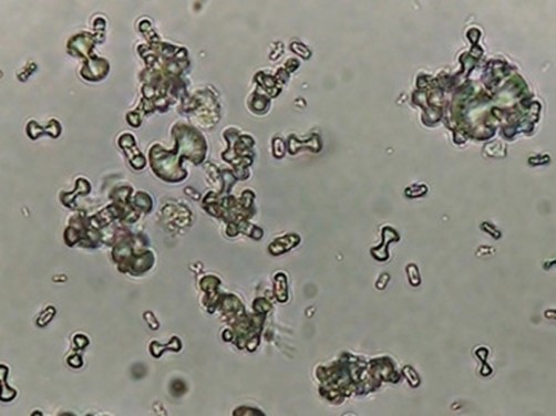
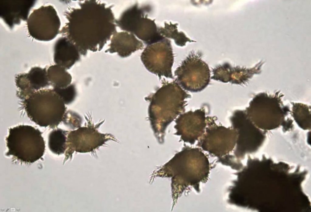
Ammonium Biurate
Although these are one of the strangest looking crystals, their appearance holds little to no clinical significance. Their presence typically signifies a urine sample that is old or improperly preserved. It may be wise to suggest a recollection of the sample if these crystals are seen in order to perform an accurate urinalysis on a fresh sample. These crystals are described as having a “thorny apple” appearance.
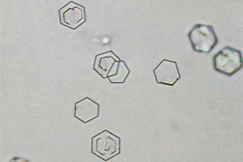
Cystine
Cystine crystals are quite rare, as cystine is normally reabsorbed from the kidneys. However, there is a recessive genetic condition that disrupts this process and causes cystine to build up in the urine, leading to the formation of these crystals. People with cystinuria are at increased risk for kidney stone formation as a result. Cystine crystals are flat, colorless hexagons.
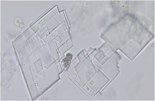
Cholesterol
Cholesterol crystals may form in neutral or acidic urine and are more likely to appear if a sample has been refrigerated. The presence of these crystals signifies renal tubular disease, and they will often be found alongside lipid drops, fatty casts, or oval fat bodies, as well as severe proteinuria. These crystals are described as flat, rectangular plates or “glass plates,” and will have notched corners.
I would like to say that this is a comprehensive list of every known urine crystal, but unfortunately, it is not! This is just a list of the ones I have seen in my first year as a Medical Laboratory Scientist. Thankfully, my lab has brightfield, phase, and polarized microscopes to examine crystal morphology. With this information, as well as the urine pH, patient condition, and various laboratory resources (including the veteran lab techs!) identifying urine crystals can be a fun investigation. Happy crystal hunting!